Carotid Ultrasound: Understanding Anatomy and Hemodynamics
- Shemein Samuda
- Sep 21, 2025
- 2 min read

Carotid artery ultrasound is a core vascular exam for detecting conditions that can lead to stroke and other serious complications. For sonographers, mastering both anatomy and hemodynamics of the carotid system is critical for accurate diagnosis and patient safety.
Why Carotid Ultrasound Matters
Stroke prevention: Detects narrowing or blockages that increase stroke risk.
Monitoring disease: Tracks progression of atherosclerosis and plaque formation.
Evaluating symptoms: Helps assess transient ischemic attacks (TIAs) or other neurological changes.
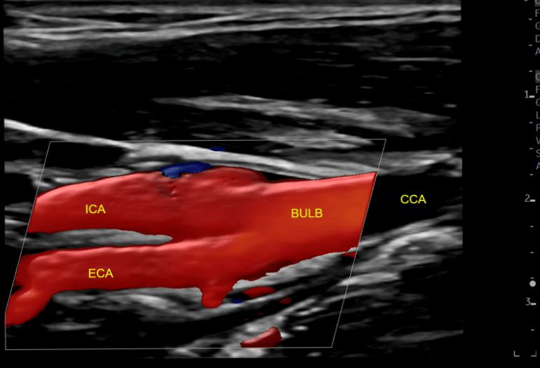
Key Carotid Artery Anatomy
Understanding the vascular anatomy the first step to precise scanning:
Common Carotid Artery (CCA): Divides into the internal and external carotid arteries.
Internal Carotid Artery (ICA): Supplies blood to the brain; typically larger, with no neck branches.
External Carotid Artery (ECA): Feeds the face and scalp; identifiable by side branches.
Vertebral Arteries: Travel through the cervical spine to join and form the basilar artery.
Tip: Correctly distinguishing ICA from ECA is one of the key to accurate doppler interpretation.
Hemodynamics and Doppler Clues
A good sonographer goes beyond structure and reads blood-flow patterns:
Normal ICA flow: Low-resistance waveform with continuous forward diastolic flow.
Normal ECA flow: High-resistance waveform with brisk systolic upstroke and low diastolic flow.
Stenosis: Elevated peak systolic velocities, spectral broadening, and post-stenotic turbulence.
Vertebral arteries: Antegrade flow is normal; reversal may signal subclavian steal syndrome.
Accurate angle correction (≤60°) is vital for accurate velocity measurements.
Carotid Ultrasound Scanning Protocol
Patient prep: Supine position, head turned slightly away.
Longitudinal & transverse grayscale imaging: Assess vessel wall and plaque.
Color Doppler: Visualize flow, turbulence, or occlusion.
Pulsed-wave Doppler: Record velocities in proximal, mid, and distal segments of CCA and ICA. Record velocities ECA and vertebral arteries.
Documentation: Save representative images with velocity measurements and any pathology.
Common Pitfalls and How to Avoid Them
Incorrect angle correction leading to velocity overestimation
Confusing ICA and ECA (check for branches and waveform differences)
Over-gaining color Doppler, which can mimic turbulence
Skipping vertebral artery assessment
Master the Technique with Hands-On Training
While guidelines are important, real expertise comes from supervised practice. At SonoSavvy Institute, our vascular ultrasound face to face training covers:
Hands-on detailed carotid scanning
Doppler interpretation and case-based learning
Strategies for consistent, reproducible results
👉 Book your private vascular ultrasound training and elevate your vascular imaging skills.

This article provides an excellent overview of carotid ultrasound anatomy and Doppler principles, emphasizing the importance of proper technique for accurate vascular assessment. High-quality diagnostic imaging plays a vital role in supporting timely clinical decisions. For patients seeking trusted MRI services in Richmond, choosing an experienced imaging center ensures advanced technology, accurate results, and compassionate care throughout the diagnostic process.